


13. Case of the month: chest pain
Case and images James Dent A 44F presents with chest pain for 20 mins, central chest reflux/burning sensation. Onset on leaning forward to open drawer. Now just in left shoulder. Then tingling to bilateral hands, feet and generalised and spasming of hands. Recently well OE: eyes closed, SaO2 100% on…
Read More >12. Case of the month: Renal colic
Case and images Sarah Vaughan Discussion Genevieve Carbonatto A 55 year old man presents to the ED with 5 days of loin to groin pain and microscopic haematuria. He was started on nitrofurantoin as an outpatient for a possible UTI. A point of care renal ultrasound is performed This is…
Read More >11. Case of the month: Right hip pain
Images Daniel Loui Text Genevieve Carbonatto A 60 year old lady presents with recurrent syncopes associated with frequent falls. 2 weeks prior to presentation she had a fall on her right buttock causing right hip pain. Subsequently she developed a small heamatoma over her right buttock and a larger right…
Read More >10. Case of the month: Stanford type B dissection
Images Earl Butler, text Genevieve Carbonatto A 55 year old lady presents with acute numbness to her right leg and "dizziness" lasting a few hours. No back pain. No chest pain. There are no other neurological symptoms. BP 175/85 HR 72/min, RR 16/min saturations 96% on room air. Because of…
Read More >9. Case of the month: PoCUS in pneumomediastinum
Images Chris Fox A 14 year old boy presents to ED with throat and neck pain after persistent vomiting. He states he vomited > 30 times over the previous 8 hours. On examination: oxygen saturation 98% on RA, BP 128/89, HR 66/min. No obvious crepitaions to the neck. A chest…
Read More >8. Case of the month: Assessment of LV function
This month I thought it might be good to review the basics of assessment of LV function. This is a PLAX. What are the 3 ways you would assess systolic LVF in this patient? To help you with this, compare the clip with the PLAX of a patient with normal…
Read More >7. Case of the month: FELS (focused ECHO in life support) in patient with dyspnea
(Case and images courtesy of Dr Ed Brentnall ultrasound reg, text Jay Perera, Genevieve Carbonatto) Triage: -3.06pm on Wednesday afternoon -Cat 3 to acute bed. 2/52 of worsening SOB on exertion +dry cough. Postural drop with CDA. -Observations: BP 116/65, HR 80, RR 24, sats 96% on room air, temp…
Read More >6. Case of the month: AAA
Images and case Danielle Martinez An 89 year old lady presented to hospital with severe abdominal pain and collapse on a background of a known AAA. On arrival, she was hypotensive and pale with a reduced level of consciousness. A Point of Care Ultrasound was performed. A shows a transverse view…
Read More >5. Case of the month: Pneumonia
Images Dr Ed Brentnall. History: A 22 year old man presents to ED with 1 week of a sore throat, a mild productive cough, fevers and rigors for 8 hours. On examination : temperature 39.6, HR 143/min, RR 28/min, BP 133/60, Sats 98% on RA Bloods were ordered. His WCC…
Read More >4. Case of the month: Ectopic pregnancy
Images Micky Fiorentino Triage: A 46 year old lady presented to the Emergency department after being referred by the early pregnancy assessment clinic for an ectopic pregnancy and admission under O and G. She is 11 weeks pregnant. It is an IVF pregnancy. She is hemodynamically stable and is not complaining…
Read More >3. Case of the month: Gallstones, dilated CBD
Case and images courtesy of Dr Micky Fiorentino (ultrasound under supervision of Dr James Dent) Triage at 1pm: 46F, representation with recurrent RUQ pain. Known gallstones. Representing with further pain, yellow skin colour and pale stool and dark urine. O/E Minimally tender RUQ, Murphy’s negative Jaundiced Initial blood tests: Hb…
Read More >
Eustachian valve
A 72 year old lady presented to the Emergency Department with syncope. A point of care ECHO was performed. What is the structure in the right atrium? This is a Eustachian valve. The differential diagnosis would be a tumour, a thrombus or a vegetation. In the context of syncope…
Read More >2. Case of the month: FELS Right heart failure
Dr Chris Harrington (FACEM) presents 2 cases for this month. Both demonstrate the importance of ECHO in the context of the deteriorating patient with heart failure. A 48F is referred to the Emergency department by her cardiologist with exacerbation of right heart failure and rapid AF PHx aortic valve repair…
Read More >1. Case of the month: Focussed Echo in Life Support(FELS)
(Case and images courtesy of Jay Perera, text Jay Perera and Genevieve Carbonatto) Triage: 30F presents with 2/7 of worsening palpitations, worse post Salbutamol. She has a history of PTSD and anxiety. HR 78 regular. Further history: 3 years of fatigue and lethargy Complains of intermittent palpitations and dyspnoea. Trialled…
Read More >
Pseudoaneurysm
Images James Dent An 80 year old had an unsuccessful cannulation for a CT scan. 4 days later she presented with bruising and tenderness of the left antecubital fossa and a palpable pulsating mass. This is the US of the area B mode sonography shows a large pseudoaneurysm . The…
Read More >
Pitfall – Stones in the gall bladder – Adenomyomatosis
Images Ying Ying Lee, text Genevieve Carbonatto A 44 year old man presents with acute right upper quadrant pain. A point of care ultrasound is performed to exclude a biliary cause. While scanning the patient in the supine position, stones are visible in the body of the GB which do…
Read More >
Lung Ultrasound findings in COVID 19
Text Genevieve Carbonatto Specific examination ultrasound findings in COVID 19 are not unique to the disease. Lung findings include pleural line irregularities, pleural thickening, B lines and consolidations. What is striking is the remarkably similar distribution of lung pathology from patient to patient. The posterolateral areas are generally affected first…
Read More >
Features of lung ultrasound in COVID 19 infection
Text Genevieve Carbonatto Literature is coming out on the lung ultrasonography of novel coronavirus. This is a summary of the literature so far The features of lung ultrasound are not specific for COVID 19 pneumonitis or pneumonia but highly suggestive in patients presenting with a history suggestive of infection with…
Read More >
Causes of Aortic Stenosis
Kathryn Statham Aortic stenosis It is the most common valvular heart disease in developed countries It's prevalence is increasing with our ageing population It is found in 3% of patients > 75 yrs It is the most common valve disease requiring surgical intervention in developed countries. TAVI is increasingly the…
Read More >
Assessment of aortic regurgitation
Kathryn Statham, Genevieve Carbonatto AR is rarely physiological. If it is present then a cause needs to be found. ECHO is used to determine Aetiology Estimate severity Assess chronicity This is done first by imaging the heart with 2D echo and looking at: valves aortic root size LV cavity size…
Read More >
Aortopathy – Aortic root dilatation
Images Ava Ghalini text Genevieve Carbonatto A patient is referred by his GP to the Emergency Department for episodes of exertional upper chest pain over a period of 2 weeks. A troponin organised by his GP was 28. On arrival he is asymptomatic. BP 180/80, HR 100/min. A systolic and…
Read More >
Ruptured ectopic, pseudosac
Images Bashir Chakar, text Genevieve Carbonatto A 36 year old lady presents with sudden onset of right lower quadrant pain lasting 20 minutes. One hour later she experiences further abdominal pain prompting her presentation to the Emergency Department. She has had 2 positive home pregnancy tests. On examination she is…
Read More >
Heterotopic pregnancy
Images Evy Panos, Text Genevieve Carbonatto A 32 year old lady presents by ambulance to the Emergency Department in shock. She describes having felt dizzy but not unwell and then suddenly collapsed. She remembers being escorted in a wheelchair into an ambulance. She is 19 weeks pregnant and has had…
Read More >
Hydronephrosis and aberrant renal artery
Images Evy Panos A 32 year old man presents with right loin pain after a drinking binge. His friends have urged him to attend the Emergency Department because he always complains of right loin pain after drinking in their company and they would like him to get to the bottom…
Read More >
Pulmonary Embolism – Right ventricular thrombus
Images Nick Stewart text Genevieve Carbonatto A 22 year old patient presents to the Emergency Department with calf pain. On further questioning she has had a presyncopal event the day of presentation and complains of shortness of breath. She has recently been put on tranexamic acid for heavy menstrual bleeding.…
Read More >
Cardiac tumour
Images Roger Burrell Text Genevieve Carbonatto A 60 year old man presented to the Emergency Department short of breath having a known right ventricular cardiac tumour with metastases to the lung, liver, spleen and bone. On examination saturating 100% on RA, bilateral pitting pedal oedema, BP, 120/60, HR 68/min. An…
Read More >
Echocardiography during cardiac arrest: COACHRED
Text Genevieve Carbonatto The management of cardiac arrest has been intensively studied over the past 20 year and other than early and good quality CPR and early defibrillation in VF, very little has shown to improve the prognosis in cardiac arrest. The last 20 years has shown that high dose…
Read More >
Pulmonary hypertension in a child
Images Juan Chiang A 13 year old girl presents to the Emergency Department after a syncopal event at school. This was not associated with prior chest pain or SOB. This is her first syncopal event. ECG Chest Xray A point of care ultrasound is performed. The following clip shows…
Read More >
VSD in a 3 month old
Images Hanaho Imamura, text Genevieve Carbonatto A 3 month old girl is brought into the Emergency department. Her parents state that she has become more unsettled recently, not feeding as well and appearing to be more short of breath with feeding. She is not settling with feeding, requiring to be…
Read More >
Coarctation of the aorta in a neonate
Images Chris Harrington, text Genevieve Carbonatto A 11 day old presents to the Emergency department in acute respiratory distress. His mother states that his SOB has increased markedly over the past 24 hours. While on the way to hospital he has an episode of tachypnea cyanosis and not breathing for…
Read More >Stanford type B dissection
Images Earl Butler, text Genevieve Carbonatto A 55 year old lady presents with acute numbness to her right leg and "dizziness" lasting a few hours. No back pain. No chest pain. There are no other neurological symptoms. BP 175/85 HR 72/min, RR 16/min saturations 96% on room air. Because of…
Read More >
Bacterial endocarditis : Mitral valve
Images Nick Stewart, text Genevieve Carbonatto A 50 year old man presents to the ED with a 6 week history of general malaise, fevers, night sweats and weight loss (10 kg). He had been prescribed 7 courses of Augmentin by his GP and felt generally better on this antibiotic. An…
Read More >
Diverticulitis
Images and Text Genevieve Carbonatto A 45 year old man presents with a 3 day history of diarrhea and increasing LIF pain. He is afebrile. His diarrhea has tapered off. On examination he is tender in his LIF. A point of care ultrasound is performed using a curvilinear probe. He…
Read More >
Dissection
Images Dr Juan Chiang An 85 year old man is brought in by ambulance after a syncopal event. He was hypotensive on scene, BP 78/50. He is given fluid by the paramedics but remains hypotensive. He has no chest pain and is not short of breath. On arrival at the…
Read More >
Lung ultrasound for pulmonary embolism
Images Edward Christian Text Genevieve Carbonatto A 65 year old man presents to the Emergency Department with a 1 week of cough and left sided chest pain. The pain is intermittent but worse at night when lying down. It is not exacerbated by activity. The patient is experiencing shortness of…
Read More >
Submassive Pulmonary Embolism – a critical care ECHO skill?
Images and Text Genevieve Carbonatto If we consider that there are different levels of expertise in the acquisition of knowledge of echocardiography for the Critical Care Physician, the recognition of acute cor pulmonale which we see with submassive pulmonary embolus has been described as a Level II skill, level I…
Read More >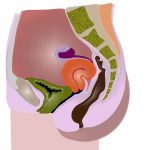
Pitfall : Retroverted uterus
Images and text Genevieve Carbonatto A 32 year old lady presents with PV spotting. She is thought to be 8 weeks pregnant. A point of care transabdominal scan is performed in the Emergency Department. This is her transverse scan of the pelvis The bladder is empty. There appears to be…
Read More >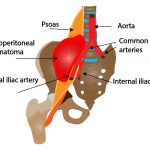
Giant retroperitoneal haematoma
Images and text Genevieve Carbonatto An 88 year old woman presents to ED from a nursing home hypotensive. She has recently been admitted to hospital with pneumonia, CCF, AF and bilateral below knee DVT's. She was discharged on warfarin. She is generally feeling tired, has abdominal pain and is vomiting…
Read More >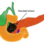
Dilated CBD, pancreatic carcinoma
Images and text Kezia Mansfield and Olga Gaitsgory An 84 year old woman presents to the emergency department with a week of nausea, anorexia and general malaise, without abdominal pain. She has a past history insulin dependent diabetes, GORD and osteoarthritis. It was noted on examination that she was moderately jaundiced,…
Read More >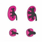
Acute vs Chronic Hydronephrosis
Text and Images Oli Gaitsgory and Kezia Mansfield Clinical presentation: A 70 year old man with a background of metastatic colorectal cancer presents to the Emergency Department. He has a known large pelvic mass causing L ureteric obstruction, with a L ureteric stent in situ. He presents with 1 week of…
Read More >
Eyeballing LV function : Test yourself
Gollum, Wellington Airport, Weta Studios Estimating ejection fraction (EF) can be done by eyeballing LV contraction on ECHO. This requires some skill and expertise. A very basic assessment in the Emergency department requires a parasternal long axis (PLAX), a parasternal short axis (PSAX) and a 4 chamber view (4CV). What…
Read More >
Cholecystitis and obstruction of the CBD
Images Victoria Bond Text Genevieve Carbonatto A 42 year old lady presents to the Emergency Department with RUQ pain. She has had episodes of cholelithiasis in the past. She is mildly tender in her RUQ. A point of care ultrasound is performed along with the history and examination. This is…
Read More >
Renal stones in PUJ
Images Chris Harrington, Text Genevieve Carbonatto A 60 year old man presents with right renal flank pain radiating to the groin. He was known to have a 6mm stone in his VUJ from a CT scan a few months back. At the time he had no hydronephrosis. A point of…
Read More >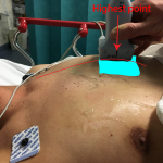
Pneumothorax : Where do we place the probe?
A 56 year old man presents to the Emergency department from another hospital for trauma assessment. Earlier that morning he had fallen off a roof and injured his chest and left hand. He has had 3 chest ultrasounds performed by 3 different operators and a pneumothorax was missed. It was…
Read More >
Trauma: Right kidney laceration
Images Stuart Napier, text Genevieve Carbonatto A 24 year old presents to the emergency department with right upper quadrant pain after playing rugby. He tripped, landing on his right elbow jamming into his right upper quadrant. He was immediately winded and felt right upper abdominal pain. On waking the next…
Read More >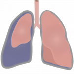
Pneumothorax : Test yourself
We have added lung scans of patients with and without pneumothoraces. Test yourself! Pneumothorax/No pneumothorax 1. 2. 3. 4. 5. \ 6. 7. 8. 9. 10.
Read More >
Intussusception in a 6 year old
Images and text Genevieve Carbonatto A 6 year old girl presents to the Emergency department with dysuria and abdominal pain. The symptoms have been intermittent until the morning of the presentation when the symptoms were constant and also associated with a left sided limp. She is unwilling to weight bear…
Read More >
Bacterial endocarditis : Tricuspid valve
Images Sarah Vaughan, Text Genevieve Carbonatto A BAT call arrives. A 42 year old man recently discharged against medical advice from hospital is coming acutely short of breath (SOB). 2 weeks prior he self discharged despite acute liver failure from hepatitis C and positive blood cultures for strep mitis. He…
Read More >
Pitfall: Ruptured cornual ectopic pregnancy
Images Nick Sidler, text Genevieve Carbonatto A 32 year old woman presents to the ED with her husband and 5.00 am. The woman is clearly unwell. She is directed straight to the resuscitation bay. Her husband tells you that shortly before 4.00 am she awoke with severe acute abdominal pain.…
Read More >
ET tube malposition
Images and text Genevieve Carbonatto. You get a BAT call. A 25-year-old with acute asthma has had a cardiopulmonary arrest. He has been intubated by the paramedics and received 1 mg of adrenaline with return of circulation. He is arriving in 5 minutes. A resus team is organised and you…
Read More >
Trauma : knife through chest
Ultrasound images and text Genevieve Carbonatto A 53 year old lady presents to the Emergency Department after having stabbed herself in the chest in a suicide attempt. The knife is still in the chest close to and to the left of the sternum in the 5th intercostal space. She is…
Read More >
Pericardial Tamponade
Images : Sarah Vaughan, text Genevieve Carbonatto A 56 year old man presents to the Emergency department short of breath and unwell. He gives a history of 2 hours of shortness of breath and chest pain. He has been recently diagnosed with metastatic adenocarcinoma. These are his vital signs He…
Read More >
Oesophageal intubation : Double tract sign
Images and Text Genevieve Carbonatto Tracheal intubation Ultrasound can be used as an adjunct to directly visualising endotracheal tube placement. The linear probe is placed in the transverse position with the index marker pointing to the right on the neck above the sternal notch. The structures are identified. These include…
Read More >
Alcoholic cirrhosis
Images and text Genevieve Carbonatto A 54 year old man presents to the Emergency Department with abdominal pain and jaundice. He has a distended abdomen. He is known to have alcoholic cirrhosis. A point of care ultrasound was performed. The following demonstrates some of the basic features of cirrhosis with…
Read More >
LV Aneurysm
Images and text Genevieve Carbonatto A 56 year old man presents to the Emergency Department with chest pain. This is his ECG The ECG shows deep Q waves and ST elevation in his anteroseptal leads These are his vital signs: BP 120/69 mmHg, HR 89/min, Sats 100% on RA, RR16/min…
Read More >
Cavernous haemangioma
Images and text Genevieve Carbonatto An abdominal ultrasound is performed on a young woman for right upper quadrant pain. This is what is seen while scanning through the liver This is a cavernous haemangioma. It looks like a snowball on ultrasound. Characteristics of haemangiomas: Blood filled cavernous space lined with…
Read More >
Right ventricular thrombus
Images and text Genevieve Carbonatto A 35 year old man presents to the Emergency Department with severe shortness of breath. He is known to have an amphetamine induced cardiomyopathy. His heart rate is 129/min, BP 116/76. He has mild pedal oedema. His JVP is elevated and on auscultation he has…
Read More >
Emphysematous kidney
Images and text Genevieve Carbonatto A 55 year old man with bilateral renal transplants and diabetes presents to the Emergency department with a 6 day history of right upper quadrant pain and severe back pain, fever, diarrhea, nausea and vomiting. On examination his HR is 95/min. He has a systolic…
Read More >
Pitfall: Blood clots and ruptured ectopic pregnancy
Images and text Genevieve Carbonatto It is a busy Thursday evening in the Emergency Department. There is no place to put patients and assess them. The only bay which may be available is occupied by a lady who refuses to leave the bay because she feels so ill. You call…
Read More >
Pitfall: Painless ruptured ectopic pregnancy
Images Mark Russel, text Genevieve Carbonatto A 35 year old women presents to the Emergency Department for a review of a possible ectopic pregnancy. She is 7 weeks pregnant by dates. An outpatient scan was performed the day before for painless vaginal bleeding . It did not show an intrauterine…
Read More >
Assessment of Pericardial effusion
Text and images Genevieve Carbonatto Pericardial effusions are commonplace. A pericardial effusion is not synonomous with pericardial tamponade. Pericardial tamponade is a medical emergency characterised by shock and associated with a number of clinical criteria including Becks triad (hypotension, decreased heart sounds, elevated JVP) pulsus paradoxus ( > 12 mmHg…
Read More >
Trauma – fractured ribs undiagnosed on chest Xray
Images Roger Burrell, text Genevieve Carbonatto A 54 year old man presents one week after a fall. He is complaining of severe left lower rib pain especially with respiration. His chest Xray looking for rib fractures does not show any fractures, nor is there a pneumothorax or a pleural effusion.…
Read More >
B lines – how best to see them
Thankyou Justin Bowra for this post. Ultrasound for interstitial lung disease and pulmonary oedema Key points B lines are found in pulmonary oedema and in other interstitial syndromes B lines start at the pleural line, move with breathing, are very bright, and reach much further down the screen (more than…
Read More >
Trauma – Haemopericardium
Images Tina Cullen, text Genevieve Carbonatto It is 10.00 pm and you get a BAT call. There has been a fight at the local pub and the ambulance are bringing in a 34 year old who has been stabbed in the chest. He is tachycardic and hypotensive, GCS 14. They…
Read More >
Ice cardiomyopathy, apical thrombus
Images Sarah Vaughan, text Genevieve Carbonatto A 38 year old man presents to the Emergency department with shortness of breath which he has had for 4 weeks. His symptoms have worsened over the past 3 days. He can no longer go up 2 flights of steps to his apartment and…
Read More >
Takotsubo cardiomyopathy
Images and text Genevieve Carbonatto A 52 year old lady presents to the Emergency Department with chest pain. After an argument with her husband about his spending she developed acute severe central chest pain radiating down her right arm. She is mildly SOB. There is no past history of note,…
Read More >
Biliary Obstruction: Double Barrel sign, Monkey Puzzle sign
Images and text Genevieve Carbonatto A 73 year old man presents to the emergency department jaundiced. He says he has not been feeling unwell but that over the past 4 days the white of his eyes have turned yellow as well as the skin of his face. His urine turned…
Read More >
ECHO : image optimisation
Images and text Genevieve Carbonatto The most difficult and frustrating ultrasound examination in the Emergency department must be the ECHO exam. What can we do to optimise our images ? Here are some tips and tricks. 1. Positioning the patient Some of our patients in the Emergency Department cannot be…
Read More >
Type A dissection
Images Bashir Antoine Chakar (Emergency registrar) Text Genevieve Carbonatto You get a BAT call. The ambulance are bringing in a 65 year old lady who developed acute onset chest pain radiating to her back. The paramedics tell you that she was found to be diaphoretic with a systolic BP of…
Read More >
Appendicitis
Images and text Genevieve Carbonatto A 14 year old boy presents to the Emergency Department with abdominal pain. The pain had started acutely the day before and he had been unable to sleep because of the pain. As you lead the patient to the examination room you note his antalgic…
Read More >
Normal gut ultrasound
Images and text Genevieve Carbonatto. I would like to thank the IBUS group , in no particular order, Torsten Kucharzik, Christian Masser,Giovanni Maconi, Frauke Petersen, Kim Nylund, Ruediger Goertz, Emma Calbrese, Anil Kumar Asthana, Kerri Kovak, Rune Wilkens and Stefania Carmagnola for their fantastic course on gut ultrasound which has…
Read More >
Bowel oedema
Images Bashir Antoine Chakar (Emergency Registrar) and text Genevieve Carbonatto A 72 year old man presents to the Emergency Department. He awoke in the early hours of the morning with acute abdominal pain which he thinks is due to what he had eaten the night before. He describes his pain as…
Read More >
Crohn’s disease – Bowel obstuction
Images and text Genevieve Carbonatto. A 48 year old man presents to the Emergency department with severe abdominal pain. He has Crohn's disease and frequently presents with abdominal pain. He has had a previous entero-enteric anastomosis. He is also self employed and hard working and always waits until his pain…
Read More >
Intussusception: Left upper quadrant mass
Images Genevieve Carbonatto, text Genevieve Carbonatto An 80 year old lady presents to the Emergency Department with a 4 month history of feeling unwell on and off associated with a 4 kg weight loss during that time. She presented acutely because the night before she developed mild abdominal pain and…
Read More >
PFO
Images Genevieve Carbonatto and Tina Cullen. Text Genevieve Carbonatto A 35 year old lady presents to the Emergency Department with chest pain. She has had a recent admission for drainage of a pericardial effusion (non malignant) and is concerned that it may have reaccumulated. An point of care ECHO was…
Read More >
Cardiac arrest : Aortic dissection
Images Sarah Vaughan (Emergency Registrar) text Genevieve Carbonatto There is a BAT call. The ambulance are bringing a 72 year old woman who suddenly , in front of her husband, was seen to slump to one side and appeared to have a left hemiplegia. In transit to hospital she has…
Read More >
Renal colic – the twinkle artifact
Images Bashir Antoine Chakar, text Genevieve Carbonatto A 32 year old man presents with right flank pain. He had an episode of renal colic 2 years ago and presented to the Emergency Department back then. He had a CT KUB done at the time which showed a small stone in…
Read More >
Renal colic
Ultrasound images Genevieve Carbonatto and Tina Cullen text Genevieve Carbonatto A 35 year old man presents to the Emergency Department with acute onset right flank pain. He has been having "niggles" of pain in the last week but suddenly at work he develops severe pain. He has microscopic haematuria on…
Read More >
Ruptured ectopic pregnancy
Images Sarah Vaughan (Emergency Registrar) Text Genevieve Carbonatto A young 30 year old lady is brought in by ambulance with acute onset severe lower abdominal pain. The first thing you want to exclude is a ruptured ectopic. On arrival she is pale and looks shocked. She says she had a…
Read More >
Aortic Dissection – Stanford Type A
Echo images Matthew Oliver (Emergency Physician) Text Genevieve Carbonatto There is a BAT call. The ambulance are bringing in a man with severe back pain. 5 minutes later a 70 year old man is wheeled into the rescusitation room. He is in severe distress. He states he was lifting a…
Read More >
Air: subcutaneous emphysema vs pneumothorax
Images and text Genevieve Carbonatto A 66 year old presents to the Emergency Department after a fall onto her right chest wall. She had slipped in her bedroom and landed backwards onto a cabinet in her bedroom . She is in great pain on arrival to ED. This is her…
Read More >
Cardiac Amyloidosis
ECHO images and text Genevieve Carbonatto The following patient presented to the Emergency Department after a syncopal event walking up a flight of stairs. He had become more short of breath (SOB) over the past week or so, finding it hard to walk more than 50m without beocoming SOB. On…
Read More >
DVT – Why use Doppler?
Images Sarah Vaughan, text Genevieve Carbonatto A young 26 year old girl presents to the Emergency Department with excruciating pain in her left leg. She developed the pain acutely that morning and she can't walk on it at all. She helps you with your diagnosis by saying that she has…
Read More >
Cardiomyopathy – Postpartum Shortness of Breath
Images and text Genevieve Carbonatto History: A young 32 year old lady presents to the Emergency Department SOB (short of breath). She is 4 weeks postpartum and was well during her pregnancy. She did not suffer from pre eclampsia during or just after her pregnancy. The baby was born by…
Read More >
Empyema
Ultrasound images and text Genevieve Carbonatto. Featured image: Oceanographic museum Monaco. A 45 year old man presents to the Emergency Department with a 6-month history of feeling generally unwell with fevers and a chronic cough. He is no longer able to cope at home. On arrival he is saturating 90%…
Read More >
Bacterial Endocarditis – Aortic valve
Images Genevieve Carbonatto. Text Genevieve Carbonatto A young man presents to the Emergency Department with SOB (shortness of breath). He has been sent in by his flatmates because he is so unwell. They leave as soon as he presents to triage. He is an intravenous drug abuser, but says he…
Read More >
FAST – Focus on the RUQ – False +ve
Text and images Genevieve Carbonatto There is a BAT call. A young 35 year old has fallen from a 3 story building and essentially landed on his feet. He has extensive leg injuries and possible chest and abdominal injuries. BP 95/60 HR 110/min RR 25/min Saturating 96% on RA. The…
Read More >
Early Pregnancy Fetal Heart Beat – Now you see it, now you don’t
Images and text Genevieve Carbonatto PV bleeding in early pregnancy is common. This is probably one of our most common presentations to the emergency department. Transabdominal ultrasound is less sensitive in the first trimester pregnancy than transvaginal ultrasound. Seeing a fetal heart beat confirms fetal viability. It is important to…
Read More >
Internal hernia
Ultrasound images Sanjay Ramrakha. Text Genevieve Carbonatto and Katherine Bennett The Emergency department gets a pre arrival call from a GP. He is sending in a sick looking 10 year old who has been vomiting overnight. He thinks this may be gastro On arrival a pale, mottled, barely responsive child…
Read More >
Discovering the aorta through ultrasound
ECHO images Tina Cullen. Text Genevieve Carbonatto A 65 year old lady presented to the Emergency department with a 6 month history of abdominal pain. The pain was vague and mainly located in her pelvis but at times was epigastric. A bedside point of care ultrasound was performed. This was the…
Read More >
Systolic anterior motion SAM
Images Genevieve Carbonatto This gentleman presented septic to our department. He had a history of chronic renal failure requiring dyalisis. BP 88/60, HR 90/min. An ECHO was done in the ED to find a possible cardiac cause, other than hypovolaemia from sepsis, for his hypotension Watch the clip below. Watch…
Read More >
Shock – Left Ventricular Outflow Obstruction
Echo images Tina Cullen, text Genevieve Carbonatto The Emergency phone rings directly from the paramedics. They have a 59 year old woman with acute onset chest pain. BP 75/40, diaphoretic. HR 110/min in atrial fibrillation. She has a past history of hypertension. The ECG is faxed through Widespead ST…
Read More >
Giant Bulla
Images and text by Genevieve Carbonatto A 72 year old man arrived in the Emergency Department directly from a private radiology imaging service saying he had been told to present immediately to the Emergency Department for treatment of his pneumothorax. He had had a previous pneumothorax and knew all about chest…
Read More >
Incarcerated umbilical hernia
Images and text by Genevieve Carbonatto A 54 year old presents with an umbilical hernia. He says that he has always been able to push it back, but not this time. This is his ultrasound scan Note the umbilical hernial sac. The loop of bowel is not moving within the…
Read More >Right hip abscess – Pus stalsis
Images by Lee Juan Chiang A 45 year old diabetic presents with a right hip abscess. This is his ultrasound With probe compression, pus can be seen moving within the abscess. This is called 'pus-stalsis" and is diagnostic of an abscess. Discussion Skin and soft tissue abscesses account for 2%…
Read More >Deep buttock abscess.
Images by Genevieve Carbonatto A patient presented to the Emergency with a very painful right buttock. There was no area of erythema and the buttock was not enlarged . It was however intensely painful to move the right leg. To investigate what might be happening beneath the subcutaneous tissue, ultrasound…
Read More >
Gallstones in 4 month old
Images and text by Genevieve Carbonatto and Jeremy Fry Name changed to protect patient confidentiality. Sam was 4 months old when he was brought by his parents to the ED. He presented with acute onset of crying and distress which awoke him from sleep. He was grunting and drawing his knees…
Read More >
TB cardiomyopathy – All done and dusted?
Ultrasound Images and text by Genevieve Carbonatto Case History A 60 year old man presented to ED with a one day history of severe SOB on the background of a 2 month history of increasing SOB. He was so breathless that it was an effort to walk 3 to 4 metres.…
Read More >
Aortic endoleaks
Images and text by Genevieve Carbonatto An 85 year old presented to our Emergency Department with light headedness fatigue and SOB. BP 90 systolic on arrival, HR 100/min Bloods: haemoglobin of 78g/l, creatinine of 172micromol/l and urea of 13.9mmol/l. Haemoglobin 3 weeks prior to admission 90g/l. Past history of AAA endovascular…
Read More >
Intussusception – The doughnut sign or target sign, the pseudokidney sign
Images and text by Genevieve Carbonatto Case history A 42 year old man presented to the Emergency department at 4.00 am with a history of sudden onset central abdominal pain since 2.00 am. The pain was constant, in fact, it was getting worse. It was not associated with vomiting. He had…
Read More >