


Images and text Genevieve Carbonatto
A 73 year old man presents to the emergency department jaundiced. He says he has not been feeling unwell but that over the past 4 days the white of his eyes have turned yellow as well as the skin of his face. His urine turned very yellow 4 days ago and has progressively become dark. He has no pain anywhere. His GP has sent him in for investigation. He admits to being a heavy drinker. He has a past history of hypertension. On examination he is jaundiced, has palmar erythema, a soft non tender abdomen and mild hepatomegaly. He is not distressed in any way.
A point of care abdominal ultrasound focussing on the liver and the biliary system is performed to look for causes of biliary obstruction.

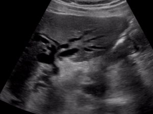
There is an apparent proliferation of vessels around the porta hepatis which are actually a mixture of very dilated bile ducts and portal veins. The bile ducts are not usually seen if they are not dilated.

This conglomeration of dilated bile ducts has been referred to as the “monkey puzzle sign”.

With colour Doppler the bile ducts can be distinguished from the vessels


With colour Doppler CBD 1.18 cm
The “double barrel sign” essentially describes 2 tubes running side by side, due to dilated bile ducts travelling along with the portal veins through the liver. Usually these bile ducts are not visible.


Left lobe of liver Right lobe of liver
The patient also has large gallstones and gallbladder wall thickening with some pericolic fluid .

Gall bladder demonstrating large round stones and thickened gallbladder wall.


Pancreas, CBD not dilated at the level of the pancreas, pancreatic duct was also not dilated
These are his blood tests: Total bilirubin 410 micromol/l, albumin 35 g/l, GGT 510U/l, ALP 510 U/l,GGT 340 U/l,ALT 155 U/l, AST120U/l, WCC 5.5 , Hb 134 g/l platelets 280
Discussion
The ultrasound in this case demonstrates biliary obstruction and a gallbladder full of stones with a thickened GB wall. The pancreas is bulky but the CBD is not dilated at that point nor is the pancreatic duct dilated. Further investigations and management include a CT scan, a formal abdominal ultrasound followed by an ERCP and stent placement and further blood tests to look for tumour markers. The cause of his jaundice was a cholangiocarcinoma and on CT and formal US he had numerous liver lesions.
What are the causes of painless biliary obstruction?
The most likely cause for painless jaundice is cancer, however inflammatory and post inflammatory causes are also a possibility.
Neoplasms
- cholangiocarcinoma
- gallbladder adenocarcinoma
- pancreatic adenocarcinoma
- metastasis
Post inflammatory
- pancreatitis
- post radiation chemotherapy
Inflammatory
- AIDS cholangiopathy
- Biliary parasites
- Primary scerosing cholangitis
Teaching point: Understand your limitations when performing any point of care ultrasound. If you are insonating the liver and you are surprised by the number of “vessels”, think about biliary obstruction and go back to basics – check the size of the CBD, look at the gallbladder, use colour to differentiate bile ducts from vessels.
Wonderful case with excellent demonstration
Thankyou Sathya. Once you see this you will never forget it and diagnosis can be made of biliary obstruction in minutes!