


Images and text Genevieve Carbonatto
PV bleeding in early pregnancy is common. This is probably one of our most common presentations to the emergency department. Transabdominal ultrasound is less sensitive in the first trimester pregnancy than transvaginal ultrasound. Seeing a fetal heart beat confirms fetal viability. It is important to record this using M mode.
The following 3 ultrasound cases will focus on this.
This is the ultrasound of a 7 week (by dates) pregnant lady.
There is an intrauterine gestational sac
By zooming in on the gestational sac, we have an intrauterine pregnancy which measures 1.08 cm (CRL)
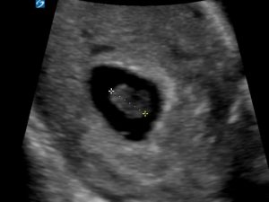
We then focus on the fetal heart
Do you see a fetal heartbeat? At times it looks like there might be one, at times you can’t see it. It is a little like the picture at the start of this post, like a visual illusion. Keep in mind that you have seen and measured a substantial fetal pole and you might be wanting very much to reassure this lady that her pregnancy is viable. There is what appears to be motion within the fetal pole, but is this motion a fetal heartbeat or is it transmitted from maternal blood flow, from her heart beat and respiration or from fluid moving within the maternal bowel? There is in fact no heartbeat and it is not possible to put an M mode through the heart to measure it. Does this mean there really isn’t one? No. All you can say is that with transabdominal ultrasound you cannot adequately display the fetal heart beat and that there is an intrauterine pregnancy but a transvaginal ultrasound will determine fetal viability.
On transvaginal scan an absent heart beat was confirmed
The following case is of 7 weeks pregnancy by dates. The patient presented with PV spotting.
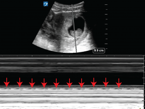
A fetal heart beat is clearly visualised. A small regular flicker can be seen in the fetal pole.

M mode enables the calculation of the heartbeat. It is 144 bpm. The M mode cursor is placed through the heart and the regular flicker of the heart beat can clearly be seen on the M mode trace.
The following patient presents with PV bleeding.
Teaching point: Be careful when confirming a heart beat in an early uterine pregnancy on a transabdominal scan. If you cannot get an adequate M mode which demonstrates the heart beating then you have not identified a heartbeat. Confirmation with a formal transvaginal scan should be organised as soon as possible.