


Images by Genevieve Carbonatto
A patient presented to the Emergency with a very painful right buttock. There was no area of erythema and the buttock was not enlarged . It was however intensely painful to move the right leg. To investigate what might be happening beneath the subcutaneous tissue, ultrasound was used.
This is a longitudinal view of his right buttock. Note the large, deep abscess. Abscess 6.47 cm X 3.08 cm


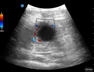
Transverse view without colour Doppler and with colour Doppler showing increased vascularity.
The abscess appears to be encapsulated by an echogenic rim and is not completely anechoic, indicating the presence of sediment or cells within the abscess. The abscess is accompanied by posterior acoustic enhancement.
Abscesses are usually readily visible below the skin with linear high frequency probes unless they are too deep as in this case. The depth of this abscess required the use of a curvilinear low frequency probe.