11. Case of the month: Right hip pain

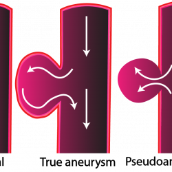
Images Daniel Loui  Text Genevieve Carbonatto A 60 year old lady presents with recurrent syncopes associated with frequent falls. 2 weeks prior to presentation she had a fall on her right buttock  causing  right hip pain. Subsequently she developed a small heamatoma over her right buttock and a larger right thigh haematoma. She presents with  Read more about 11. Case of the month: Right hip pain[…]

Pseudoaneurysm

Images James Dent An 80 year old had an unsuccessful cannulation for a CT scan. 4 days later she presented with bruising and tenderness of the left antecubital fossa  and a palpable pulsating mass. This is the US of the area B mode sonography shows a large pseudoaneurysm . The aneurysm is connected to the Read more about Pseudoaneurysm[…]

Rectus sheath haematoma

Images Genevieve Carbonatto A  65 year old man with liver failure and ascites  presents with severe acute right upper quadrant abdominal pain. Ultrasound examination over the area of tenderness reveals a heterogeneous  area in the rectus abdominal muscle – Rectus sheath haematoma The rectus sheath haematoma is the area of mixed echogenicity in the rectus muscle   Read more about Rectus sheath haematoma[…]

Right hip abscess – Pus stalsis

Images by Lee Juan Chiang A 45 year old diabetic presents with a right hip abscess. This is his ultrasound With probe compression, pus can be seen moving within the abscess. This is called ‘pus-stalsis” and is diagnostic of an abscess. Discussion Skin and soft tissue abscesses account for 2% of ED visits and the Read more about Right hip abscess – Pus stalsis[…]

Large cervical node

Images by Genevieve Carbonatto Characteristics of normal cervical nodes on ultrasound include Size < 6mm Flat or oval shape Hypoechoic echotexture Hilum is present and can be seen with colour Doppler if the node is > 5mm         Veins and efferent lymphatics  leave the node through the hilum while arteries enter the node Read more about Large cervical node[…]

Deep buttock abscess.

Images by Genevieve Carbonatto    A patient presented  to the Emergency with a very painful right buttock. There was no area of erythema and the buttock was not enlarged . It was however intensely painful to move the right leg. To investigate what might be happening beneath the subcutaneous tissue, ultrasound was used. This is a Read more about Deep buttock abscess.[…]

Infected breast sebaceous cyst

Images and text by Genevieve Carbonatto A patient presents with a small painful breast lump. This is her ultrasound The cyst is well circumscribed , is associated with posterior acoustic shadowing and has a number of internal echos suggesting sediment. Cyst measures I.48 cm X 2.31cm Discussion Features of sebaceous cysts on ultrasound (1) Located in Read more about Infected breast sebaceous cyst[…]