


Images James Dent
An 80 year old had an unsuccessful cannulation for a CT scan. 4 days later she presented with bruising and tenderness of the left antecubital fossa and a palpable pulsating mass.
This is the US of the area
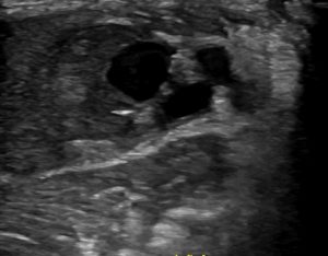
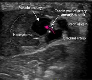
B mode sonography shows a large pseudoaneurysm . The aneurysm is connected to the artery by a small neck (between the stars).
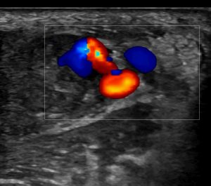
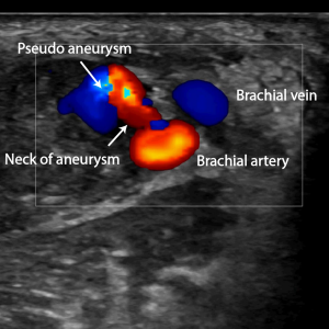
On colour Doppler flow is clearly visibly flowing from the tear in the artery, through the neck into the pseudoaneurysm. Within the pseudoaneurysm there is a typical swirling motion or colour ying yang sign.
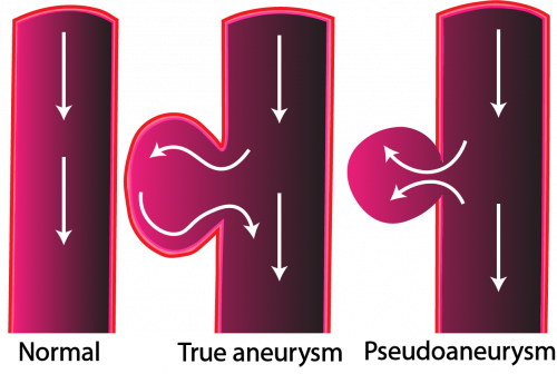
There are 2 types of aneurysms : true and false (pseudo)aneurysms.
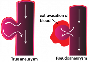
- Unlike true aneurysms, pseudoaneurysms are surrounded by haematoma, not by the vessel wall. They are in effect a contained collection of blood.
- Pseudoaneurysms are a result of blood vessel wall damage, most often iatrogenically caused by blood vessel puncture
- The incidence of pseudoaneurysms due to catheter bases interventions is between 0.6 and 4.8%. With ultrasound guided access the incidence falls to less than 0.2 %
- Femoral pseudoaneuryms after percutaneous access can occur due to (1)
– Failures of closure devices
– Laceration of the artery or branches of the common femoral artery by access needle
– Inadequate pressure or length of time holding pressure post-procedure
– Inadvertent access and/or dilation of the artery during venous procedures
– Femoral graft anastomosis can also break down over time due to suture failure, or likely more commonly due to infection of the graft material.
- They present as painful pulsatile masses
- There may be a bruit on auscultation
- thrombus that forms within the sac may embolise distally
- Ultrasound is the gold standard for diagnosis. The size, anatomy, and origin of pseudoaneurysms can be determined by ultrasound.
Treatment options
- observation if small ie less than 2 -3 cm in diameter. These may thrombose and regress.
- ultrasound guided compression.
- ultrasound guided thrombin injection. Success rate of 97% – 100% with a single treatment even while patients are on antocoagulants +/or antiplatelet therapy
- surgical repair if failure of ultrasound guided thrombin injection
Differential diagnosis
- Haematoma
- Seroma
- Infection/abscess
Reference:
- Stat Pearls Pseudoaneurysms Philip A. Rivera; Jeffery B. Dattilo https://www.ncbi.nlm.nih.gov/books/NBK542244/