


Images Bashir Chakar, text Genevieve Carbonatto
A 36 year old lady presents with sudden onset of right lower quadrant pain lasting 20 minutes. One hour later she experiences further abdominal pain prompting her presentation to the Emergency Department. She has had 2 positive home pregnancy tests. On examination she is minimally tender in her right lower quadrant. A point of care ultrasound is performed.
Longitudinal scan through the pelvis
![]()
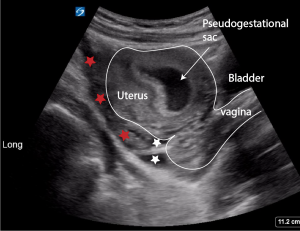
The uterus at first glance may appear to have a gestational sac. This is not a gestational sac but simply fluid within the uterine cavity called a “pseudosac” or “pseudogestational sac”. There is free fluid in the pouch of Douglas which is both coagulated (red star) and uncoagulated (white star)

The uterine cavity is filled with low level echos.
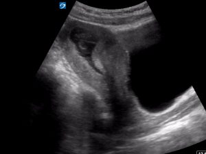
Transverse scan through the pelvis
![]()
![]()
The uterus is full of fluid. The pseudo gestational sac is surrounded by a single echogenic rim and is irregular in outline. There is no fetal pole or yolk sac present
![]()
![]()
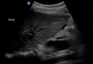
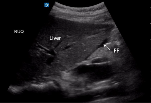
Scanning right through the pelvis reveals free fluid in the pouch of Douglas. Some is coagulated (red star) some is not (white star)
There is fluid in Morrison’s pouch and in the inferior border of the spleen and lienorenal space


The patient was transferred to theatre where a large right tubal ectopic was removed. 200 mls of clotted blood was seen in her pelvis
Discussion
The term ” pseudogestational sac” or pseudo sac” is not a sac at all but simply intrauterine cavity fluid which may confuse the scanner into thinking it is a true gestational sac in the presence of an ectopic pregnancy. It occurs in approximately 10% of ectopic pregnancies.
| Pseudogestational sac | True gestation sac |
| There is a single echogenic rim surrounding the intrauterine fluid. This can be a thick decidual reaction | A gestation sac has 2 hyperechoic rings consisting of an outer and inner ring, the decidua vera and the decidua capsularis |
| Located centrally inside the uterine cavity | Eccentrically located |
| Shape may be irregular and changes during the scan. Usually ovoid | Steady, round shape |
| Avascular | High peripheral vascular flow |
| Absence of fetal pole or yolk sac | Presence of fetal pole and fetal yolk sac |
The following clip is from a 7 week pregnant patient with a true intrauterine gestation sac. The double echogenic rim is visible as is the rounded shape of the gestational sac. A yolk sac is visible.
References
- Radiopaedia: pseudogestational sac
- Research gate : Difference between double ring sign of true gestational sac and pseudogestational sac
informative