


Images Chris Harrington, Text Genevieve Carbonatto
A 60 year old man presents with right renal flank pain radiating to the groin. He was known to have a 6mm stone in his VUJ from a CT scan a few months back. At the time he had no hydronephrosis.
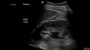
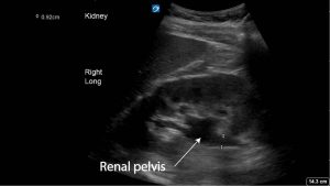
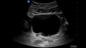
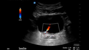
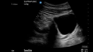
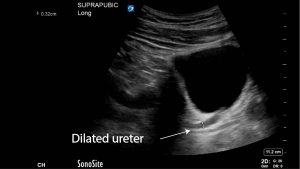
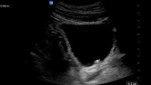
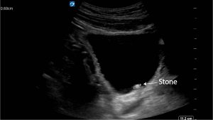
A point of care ultrasound was performed. The longitudinal view of the kidney shows mild hydronephrosis. A 6.8 mm stone is visible in the right PUJ. It is best seen in the longitudinal scan of the bladder, but visible on the transverse scan. With colour Doppler a small twinkle artefact is produced and the urinary jet is visible. The distal ureter is dilated to 3.2 mm
The patient’s pain improved with analgesia and he was discharged with Tamulosin and analgesia for outpatient urology review.
Teaching point: CTKUB remains the best imaging modality for detecting renal stones however ultrasound provides a reproducible, safe, non invasive, non radiation alternative. The European Association Urology guidelines advises that ultrasound should be the primary diagnostic imaging tool and in a situation such as this one where there is recurrence of symptoms and recent CT imaging then it is the best imaging modality.