


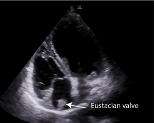
A 72 year old lady presented to the Emergency Department with syncope. A point of care ECHO was performed. What is the structure in the right atrium?
This is a Eustachian valve. The differential diagnosis would be a tumour, a thrombus or a vegetation. In the context of syncope the presence of such a prominent Eustachian valve is possibly significant as it may be a factor in causing paradoxical emboli.

The Eustachian valve is located in the right atrium. It is crescent shaped and extends from the IVC to the fossa ovalis. It is a remnant of the sinus venosus valve which directs oxygenated blood during fetal development from the placenta to the left atrium through the foramen ovale and then to the systemic circulation.
In adults, the Eustachian valve does not serve a significant physiological function and is considered a vestigial structure.
However while the Eustachian valve is not directly involved in any vital processes in adults, it can occasionally have clinical significance. In adults it is often associated with septal anomalies mainly patent foramen ovale but also ASD. It may direct blood towards a patent foramen ovale causing a persistent right to left shunt in the absence of high right sided pressures, and, in the presence of a PFO, increase the risk of paradoxical embolism.
The Eustachian valve in adults is usually a thin undulating structure visualized deep in the right atrium but occasionally, as in this case, it can be thick and quite large.
This patient did not have a PFO and previous echos showed the presence of this large Eustachian valve indicating that this structure was not a thrombus.