


Images Dr Juan Chiang
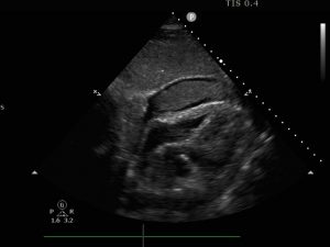
An 85 year old man is brought in by ambulance after a syncopal event. He was hypotensive on scene, BP 78/50. He is given fluid by the paramedics but remains hypotensive. He has no chest pain and is not short of breath. On arrival at the Emergency Department, he is alert, clammy, pale and profoundly hypotensive. He has a history of a thoracic aortic aneurysm. A thoracic aortic dissection is suspected.
A point of care ECHO is performed on arrival. This is his subcostal view.


Coagulated blood in the pericardial space appears hyperechoic.


In early systole (as the RA fills), The intra pericardial pressure is greater than the intra atrial pressure and there is RA free wall collapse (blue arrow). As the intrapericardial pressure increases, the RA free wall remains collapsed for longer. RA wall collapse which is greater than 1/3 of the cardiac cycle is significant.

In early diastole, as the RV is filling, if the intra pericardial pressures exceed the RV pressures, then the RV free wall collapses (blue arrow).
Unfortunately this man did not survive.
Excellent presentation and teaching