


Images and text Genevieve Carbonatto.
You get a BAT call. A 25-year-old with acute asthma has had a cardiopulmonary arrest. He has been intubated by the paramedics and received 1 mg of adrenaline with return of circulation. He is arriving in 5 minutes. A resus team is organised and you are in charge of the ultrasound on arrival. Mentally you prepare yourself for this patient. He has acute asthma. Did he have a tension pneumothorax to cause his cardiorespiratory arrest? What is his heart doing?
On arrival he is intubated, he has a HR of 110/min and a BP of 98/60. Saturations 95% on 100% oxygen. While bloods and monitoring are being organised you look at his right lung with a linear probe. There appears to be a lack of lung sliding. You cannot see any B lines or ring down artifacts. This is his M mode.

This looks suspiciously like a pneumothorax. There is one thing that bothers you however. It is this small hyperechoic “blip” on the pleural line which looks like a ring down artifact. This would exclude a pneumothorax.
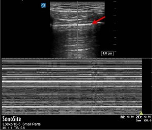
Lichtenstein has often insisted on having the B mode and the M mode side by side rather than one on top of each other. This is to marry the pleural line on the M mode with the B mode so that artifacts can be clearly interpreted in relation to the pleural line. This is what can be seen with the M mode and B mode side by side.

The pleural line is correctly identified (horizontal arrow). There are also 3 lines (vertical arrows) which end at the pleural line. This suggests a ” lung pulse” which would indicate there is no pneumothorax. A ” lung pulse” occurs when cardiac pulsations are transmitted to the pleural line and stop at the pleural line.
You examine his left chest and there is normal lung sliding, which can be visualised as the seashore sign on M mode.

A chest Xray is organised during this time and it appears that the ET tube is too far down. The team leader asks for the ET tube to be pulled back a bit. The ultrasound is repeated on the left and there is now clear lung sliding.
Discussion
Lack of lung sliding in the context of an intubated patient needs to be interpreted with caution. If there is lack of lung sliding then complications of intubation such as oesophageal intubation and right main bronchus intubation should be excluded first. The difficulty with this patient scenario is that a pneumothorax is a recognised complication of acute severe asthma and acute severe asthma may also cause lack of lung sliding. This scenario was also complicated by the fact that the lack of lung sliding was on the right, not the left, excluding right bronchial intubation suggesting left main bronchus intubation.
The “lung pulse” on M mode in this situation may be a helpful sign. A study by Lichtenstein in 2003 demonstrated that in patients with right selective intubation, a left “lung pulse” was found to have a specificity of 93% and specificity of 100% for the diagnosis of complete atalectasis before any lung changes were visible on chest Xray (1). The “lung pulse” may be easily visualised in healthy people in apnea.

What are some of the causes of lack of lung sliding other than pneumothorax?
• Right main bronchus intubation
• Oesophageal intubation
• Cardiopulmonary arrest
• Apnoea
• Pleurodesis
• Pneumonia
• ARDS
• Massive fibrosis
• Severe Acute asthma
What signs on ultrasound would indicate that a pneumothorax is not present?
- Lung sliding
- Presence of B lines
- Presence of ring down artifacts
- A lung pulse
- No “lung point” ie there is no point where the pneumothorax meets with normal lung.
Teaching point: The interpretation of lack of lung sliding in the context of cardiorespiratory arrest or in the context of an intubated patient should be interpreted with care. Check ventilation first before diagnosing a pneumothorax.
References
- Intensive Care Med. 2003 Dec;29(12):2187-2192. doi: 10.1007/s00134-003-1930-9. Epub 2003 Oct 14. The “lung pulse”: an early ultrasound sign of complete atelectasis.
Lichtenstein DA1, Lascols N1, Prin S1, Mezière G1