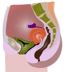
Pitfall : Retroverted uterus

Images and text Genevieve Carbonatto A 32 year old lady presents with PV spotting. She is thought to be 8 weeks pregnant. A point of care transabdominal scan is performed in the Emergency Department. This is her transverse scan of the pelvis The bladder is empty. There appears to be no gestational sac This is Read more about Pitfall : Retroverted uterus[…]

8 weeks 5 days PV bleeding

An 8 week old pregnant lady presents with 5 days of PV bleeding. A transabdominal ultrasound shows an anteverted uterus with an intrauterine gestastional sac. The fetal pole, yolk sac are visible. Yolk sac, 0.71 cm. Crown rump length 2.25 cm. There is fetal heart beat present. No sinister cause for her bleeding is seen.   Read more about 8 weeks 5 days PV bleeding[…]

Very large uterine fibroid in patient with an early intrauterine pregnancy

Images by Genevieve Carbonatto A 34 year old woman presented to the Emergency department with a at 6 weeks gestation with a palpable suprabupic mass and vaginal spotting. This is her scan.  Small gestational “squashed’ by very large fibroid Small “squashed” gestational sac with fetal pole, active HB and round yolk sac visible Focus on gestational Read more about Very large uterine fibroid in patient with an early intrauterine pregnancy[…]

Ruptured CL cyst, intrauterine pregnancy 7 weeks

  Images Genevieve Carbonatto A 7 week pregnant patient presents with acute onset abdominal pain. She has not had a scan before. Her scans show a large CL cyst, a small amount of fluid in the pouch of Douglas and an intrauterine pregancy Longitudinal view through pelvis.  Large CL cyst seen superior to bladder, small Read more about Ruptured CL cyst, intrauterine pregnancy 7 weeks[…]

6 weeks, intrauterine gestational sac

Images by Genevieve Carbonatto A patient presents to the Emergency department 6 weeks pregnant. This is her scan Longitudinal view : Intrauterine gestational sac. Fanning through the uterus in the transverse plane: Intrauterine gestational sac   Sac diameter 1.05cm. A fetal pole and heart beat should be visible if the mean gestational sac diameter is 2.5 cm Read more about 6 weeks, intrauterine gestational sac[…]

7 weeks pregnant. Intrauterine pregnancy

Images by Genevieve Carbonatt0    PV spotting. Intrauterine pregnancy. Gestational sac + yolk sac visible Gestational sac clearly intrauterine. Yolk sac and fetal pole present Round yolk sac and fetal pole with heart beat visible when fanning across the gestation sac in zoomed view Fetal HB better visualised on zoomed view of gestational sac       Read more about 7 weeks pregnant. Intrauterine pregnancy[…]