


Images by Genevieve Carbonatto
A 34 year old woman presented to the Emergency department with a at 6 weeks gestation with a palpable suprabupic mass and vaginal spotting. This is her scan.
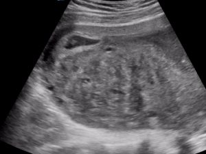

Small gestational “squashed’ by very large fibroid
Small “squashed” gestational sac with fetal pole, active HB and round yolk sac visible
Focus on gestational sac with fetal pole and yolk sac. Active HB
Discussion
Uterine fibroids are common. They occur in 20 – 30% of women over 30 years of age (1). They are usually multiple. They can be intramural (confined to the myometrium), submucosal (projecting from the uterine cavity) or subserosal (projecting from the peritoneal surface). They are oestrogen dependent.
In pregnancy they may increase in size although 1/2 will remain the same size. Some features of fibroids during pregnancy
- identified in the first trimester they are identified with a higher risk of pregnancy loss
- increased risk of pregnancy loss associated with multiple fibroids
- Large fibroids do not interfere with pregnancy or delivery unless situated in the lower uterine segment or the cervix
Ultrasound features
- Variable features
- Hypoechoic
- Heterogeneous features
- Demonstrate areas of acoustic attenuation or shadowing without a discrete mass which makes it difficult to estimate size
- Calcification
- Degeneration or necrosis
References
- Diagnostic ultrasound : Rumack