


Images Eleanor Cook and Tina Cullen
A 33 year old presents to the Emergency Department with RUQ pain. A point of care ultrasound is performed to exclude a biliary cause for her pain.
This is the transverse view of the GB. The GB could be easily missed in this view because it is full of stones. What alerts us to its presence is the shadow cast by the multiple stones within it.

The GB wall is slightly thickened at 0.34cm
The transverse view of the GB better demonstrates the gall stones . Note the confluent shadow from the numerous small stones overlying the posterior wall of the gallbladder.
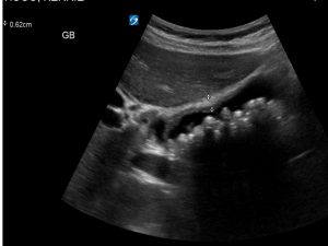
GB wall is much more thickened in this view 0.62cm

Distal CBD is dilated, 0.80 cm. The CBD should be less than 0.6 cm up to the age of 50. Normally the CBD can increase in size with age to a maximum of 0.8cm. If a patient has had a cholecystectomy, the CBD can measure up to 1 cm.
A gallstone is visible in the distal CBD. It measures 0.72 cm and casts an acoustic shadow.

Discussion
There are 2 causes of choledocholithiasis : primary and secondary. Primary choledocholithiasis occurs when stones develop within the CBD. This is relatively rare outside endemic regions (East Asia). The most common cause of choledocholithiasis occurs from migration of stones from the gall bladder.
Seeing gallstones within the common bile duct requires skill. Skill to identify small stones and skill to follow the CBD to the ampulla of Vater. In this patient, the stone is easily seen. In many cases stones, if small (<5mm) may not cast an acoustic shadow. The lateral margins of the stones may not be visible because it is compressed against the duct wall. This is not the case here. The lateral wall on the left is clearly visible because of the dilatation of the CBD. Small stones may only be visible as reproducible bright, linear echogenicity, either straight or curved.
Reference
- Diagnostic Ultrasound : Rumack