Images by Genevieve Carbonatto
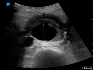
Measurement of the aorta should be made outer wall to outer wall in the transverse and longitudinal view. It is important to start as proximally as possible in the abdomen when examining the aorta. Most abdominal aortic aneurysms are infrarenal.


Proximal aorta just below the SMA is 2.32 cm and millimeters inferior to this 5.71 cm. This is best visualised in a clip
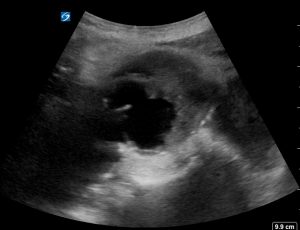
Thrombus may be seen within the lumen of the aneurysmal aorta


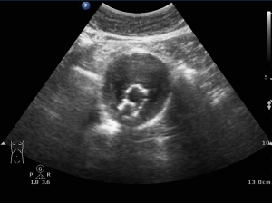
If the patient has had an aortic graft this is visualised as a hyperechoic structure within the aorta. This can be followed through to the bifurcation of the aorta. Colour Doppler should not be seen outside the graft.


Transverse view through aorta and then through graft trouser legs


Longitudinal view through graft trouser legs without and with colour Doppler


