


Physics of Ultrasound by Roger Gent

This e learning is the entire physics lecture series by Roger Gent. It covers the physical properties of ultrasound, transducer and beamforming concepts, knobology, resolution and harmonics, image artifacts, common errors in sonography caused by inappropriate technical selections or choices, Doppler – basic physics, colour, artifacts and more, bioeffects and safety and finally MI/MT phantoms.
The lectures have been split into small sections accompanied by a brief summary and some multiple choice questions. Much is also taken from his excellent book “Applied Physics and Technology of Diagnostic Ultrasound” available through ASUM.
Roger Gent has had an involvement in diagnostic ultrasound for more than 40 years, with particular interest in paediatric sonography and the physics of ultrasound. He has lectured on the physics of ultrasound for many years. He is the author or co-author of more than seventy articles, has received several awards from professional ultrasound bodies, is an Honorary Fellow of ASUM and was a member of the ASUM DMU Board of Examiners for twenty years. He was made a Member of the Order of Australia in 2009, for services to Paediatric Ultrasound.
His lectures are a must for all those sitting ultrasound exams, whether as sonographers (the DMU) or as doctors (the DDU).
Click on Show Details to view the topics in the modules below.
Modules
Lectures
- 1. The piezo electric effect
- 2. The piezo electric effect
- 3. The piezo electric effect
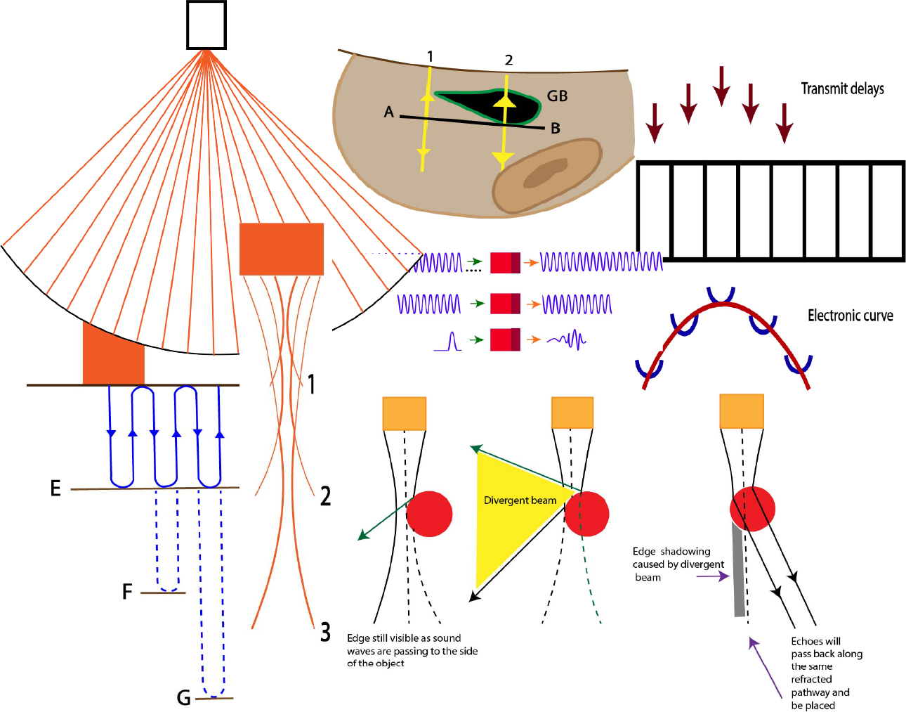
- Beam shape
- Linear array
- Curved array
- Focussing the beam : Acoustic lens
- Focussing the beam: Electronic focussing
- Matrix array
- Phased array
- Reception focussing
- Frame rate
- Multiple focal zones
- Side lobe artifacts
- The matching layer
- Resolution : spatial, temporal, contrast
- Tissue harmonics
- Completion certificate
Lectures
Lectures
- Introduction
- Acoustic shadowing
- Acoustic enhancement
- Edge artifact
- Beam width artifact
- Side lobe artifacts
- Slice thickness
- Grating lobe
- Speckle
- Reverberation
- Comet tail and ring down
- Range ambiguity
- Beam path artifacts : refraction
- Beam path artifacts: Mirror image
- Beam path artifacts: “Partial” mirrors
- Range ambiguity and electrical interference
- Artifact avoidance and minimisation
- Quiz
- Completion certificate
Lectures
Lectures
Lectures
- Case 1 : Echogenicity does not relate to density
- Case 2 : Phased array
- Case 3 : Slice thickness
- Case 4 : Curved array transducer
- Case 5 : Specular reflectors
- Case 6 : Scatterers
- Case 7 : Beam width artifact
- Case 8 : Slice thickness artifact
- Case 9 : Importance of gel for scanning
- Case 10 : Acoustic impedance mismatch
- Case 11 : Error in measurement
- Case 12 : Mirror images
- Case 13 : Reverberation artifact
- Case 14 : Cross correlation
- Case 15 : Anisotropy
- Case 16 : PRF
- Case 17 : Linear vs sector probe visualization of horizontal filaments
- Case 18 : Angle of approach (muscle)
- Case 19 : Importance of having a good window
- Case 20 : Importance of probe position. Fluid level taken from the side vs anteriorly
- Case 21 : Angle of approach (muscle)
- Case 22 : Choice of correct transducer
- Case 23 : Application of pressure to displace the bowel
- Case 24 : Lung tissue interface
- Case 25 : Angle of approach
- Case 26 : Angle of approach
- Case 27 : M mode
- Case 28 : Spatial compounding removing acoustic shadow and slice thickness (foreign body)
- Case 29 : Angle of approach
- Case 30 : Slow moving blood / angle of approach/ Doppler shift
- Case 31 : Interpretation of images (scrotum)
- Case 32 : Resistive index
- Case 33 : Mirror image
- Case 34 : Hepatisation of the lung
- Case 35 : Angle of approach (PV and CBD)
- Case 36 : Mirror image (SMA/Aorta)
- Case 37 : Colour Doppler and artifact
- Case 38 : Bioeffects Radiation force of ultrasound (scrotum)
- Case 39 : Aliasing
- Case 40 : Spectral Doppler High PRF
- Completion certificate