


Images and text Genevieve Carbonatto. I would like to thank the IBUS group , in no particular order, Torsten Kucharzik, Christian Masser,Giovanni Maconi, Frauke Petersen, Kim Nylund, Ruediger Goertz, Emma Calbrese, Anil Kumar Asthana, Kerri Kovak, Rune Wilkens and Stefania Carmagnola for their fantastic course on gut ultrasound which has spurred the following post and no doubt more to come
A recent study by Bein et al (1) showed that 812 patients presented to Emergency Departments on average across NSW (Australia) per day in 2014 . This number appears to be climbing ( 905 a day in Nov ’14) . Abdominal / gastrointestinal presentations account for around 12 % of all ED presentations (3) The ability to use ultrasound in the context of abdominal pain, is in my opinion, very rewarding. The probe is capable of directly insonating the location of a patient’s pain. Very little attention is given to the gastrointestinal tract in Emergency Medicine ultrasound and yet appendicitis, diverticulitis , acute inflammatory bowel disease and bowel obstruction are every day Emergency Department presentations. Ultrasound can provide an excellent, non radiating alternative to CT.
What does normal gut look like on ultrasound?
 Reference: Kim Nylund
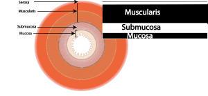
On ultrasound the 4 layers have distinct echogenicity. These alternate between hyperechoic and hypoechoic.The final layer is the mucosal interface in the centre which appears hyperechoic.


1 Serosa, hyperechoic, 2 Muscularis propria, hypoechoic, 3 Submucosa, hyperechoic 4 Mucosa, hypoechoic 5 Mucosal interface Hyperechoic. Bowel wall thickness is 2.1cm.
This is the clip. The distinct layers of the gut can be seen throughout the bowel. Just below the abdominal wall, the bowel can be seen sliding and moving and distinct layers are visible. The 5 layers as described are referred to as the gut signature and indicates normal bowel.
What probe do you use?
Depends on body habitus but in general
- Low frequency curvilinear probe for abdomen
- High frequency probe for interrogation of small gut
What are the features of small bowel?
- Presence of peristalsis – which is forward motion of bowel content not a “to and fro” of gut content, the latter suggests a bowel obstruction.
- Good visualisation of the gut wall if the body habitus allows this and the probe frequency is between 5 and 9 MHz
- In general – no air within the lumen of the small bowel.
What is be the normal thickness of the bowel wall?
- less than 3mm
- Measurement is made from the hyperechoic mucosal luminal interface to the outer edge of the hypoechoic mucosa
What are the features of large bowel?
- Little peristalisis
- Air “like clouds” within the lumen unless the bowel is collapsed
- Haustra
References
- Emerg Med Austr doi: 10.1111/1742-6723.12712 ORIGINAL RESEARCH Feeling the HEAT: Using Hourly Emergency Activity Tracking to demonstrate a novel method of describing activity and patient flow Kendall J BEIN,1 Saartje BERENDSEN RUSSELL,1,2 David MUSCATELLO,3 Dane CHALKLEY,1 Rebecca IVERS4,5 and Michael M DINH1,
- Rumack Diagnostic Ultrasound Chapter 8
- Destiny trials – unpublished data Bein et al
- Interbational ultrasound group : Module 1
Very informative post