


Text Genevieve Carbonatto
Literature is coming out on the lung ultrasonography of novel coronavirus. This is a summary of the literature so far
- The features of lung ultrasound are not specific for COVID 19 pneumonitis or pneumonia but highly suggestive in patients presenting with a history suggestive of infection with novel coronavirus
- Lung ultrasound is strongly recommended for the early diagnosis of COVID -19 pneumonia in all the patients who present to the emergency department with flu-like symptoms in novel COVID-19 era.
- Lung ultrasound has been used as a triage tool in the Emergency Department to identify those patients requiring admission and those patients able to be discharged home
- Lung ultrasound is more sensitive than chest x-ray, with a diffuse B-line pattern correlating to good response to PEEP
- Lung ultrasound patterns range from mild alveolar patterns, to severe bilateral interstitial pattern to lung consolidation corresponding to less severe to more severe disease
- Lung findings are more prominent in the posterior lower lung zones
- The spread of the infection appears to be from the peripheries of the lung to the centre of the lung in most cases
- Lung findings are more similar to pulmonary oedema than ARDS (hence recruitment manoeuvres in ICU – lying the patient prone)
- Lung ultrasound patterns also depend on comorbidities
- CT is necessary as expected to identify lesions deep to the pleural surface
- From an ICU perspective, lung ultrasound has been used instead of X Ray or CT to track the evolution of the disease, to monitor lung recruitment manoeuvres, to guide response to prone position, to manage ECMO and to make decisions on weaning the patient from ventilatory support
Other findings
- Myocarditis has been thought to be caused by COVID 19, however at present there is little evidence (histologically) that the virus affects the heart directly. Heart failure is most likely a product of the SIRS response.
- The troponin leak found in patients with COVID 19 pneumonia may also be due to the SIRS response rather than to myocarditis or myocardial infarction.
- There have been unexplained incidences of development of right ventricular failure with low pulmonary pressures in patients in the ICU
Qian-yi Peng et al ( Findings of lung ultrasonography of novel corona virus pneumonia during the 2019–2020 epidemic Qian-Yi Peng, Xiao-Ting Wang, Li-Na Zhang & Chinese Critical Care Ultrasound Study Group (CCUSG) Intensive Care Medicine (2020)) used a 12 zone method to look for lung findings (2)
The 12 zone method involves dividing the chest into 6 zones demarcated by the anterior axillary and posterior axillary lines and then into upper and basal regions. This is repeated for the other side. 2 sides therefore 12 zones.


Annals of the American Thoracic Society Volume 10, Issue 6 | December 2013
They concluded that the characteristic findings included
- Thickening of the pleural line with pleural line irregularity;
- B lines in a variety of patterns including focal, multifocal, and confluent;
- Consolidations in a variety of patterns including multifocal small, non-translobar, and translobar with occasional mobile air bronchograms;
- Appearance of A lines during recovery phase
- Pleural effusions are uncommon.
They compared lung CT with lung ultrasound features in the table below
| Lung CT | Lung ultrasound |
|---|---|
| Thickened pleura | Thickened pleural line |
| Ground glass shadow and effusion | B lines (multifocal, discrete, or confluent) |
| Pulmonary infiltrating shadow | Confluent B lines |
| Subpleural consolidation | Small (centomeric) consolidations) |
| Translobar consolidation | Both non-translobar and translobar consolidation |
| Pleural effusion is rare. | Pleural effusion is rare |
| More than two lobes affected | Multilobar distribution of abnormalities |
| Negative or atypical in lung CT images in the super-early stage, then diffuse scattered or ground glass shadow with the progress of the disease, further lung consolidation | Focal B lines is the main feature in the early stage and in mild infection; alveolar interstitial syndrome is the main feature in the progressive stage and in critically ill patients; A lines can be found in the convalescence; pleural line thickening with uneven B lines can be seen in patients with pulmonary fibrosis |
The following are examples provided from that article

Typical lung ultrasonography images of nCoV pneumonia. A. B lines; B. confluent B lines; C. small consolidations; D. translobar consolidation. (TIFF 34299 kb)
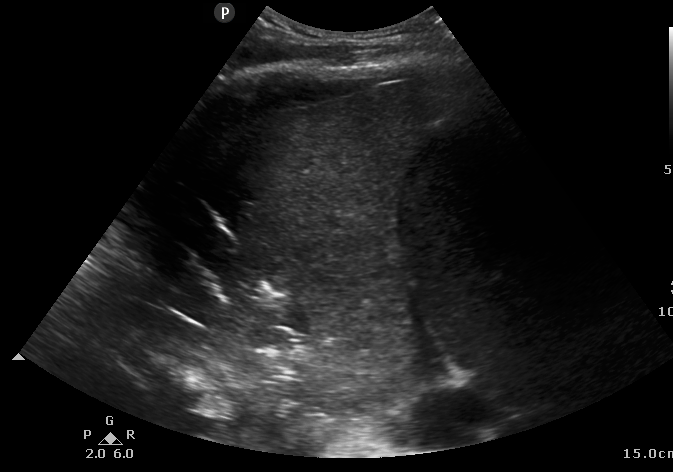
Here are some features we should be looking for which are not from Covid 19 patients (except one example) but which are examples of pathology described
- Thickening of the pleura and irregularity of the pleural line. The pleural line normally should be straight and thin.


2. . Pleural thickening with B lines (Patient with Covid 19)
B lines
3. Multifocal small consolidation/non lobar/lobar consolidation
Small consolidation


Non lobar consolidation


Another example of large consolidation
Lobar consolidation

References
- Findings of lung ultrasonography of novel corona virus pneumonia during the 2019–2020 epidemic Qian-Yi Peng, Xiao-Ting Wang, Li-Na Zhang & Chinese Critical Care Ultrasound Study Group (CCUSG) Intensive Care Medicine (2020)
- Annals of the American Thoracic Society Table of Contents Volume 10, Issue 6 | December 2013
- Huang et al. BMC Pulmonary Medicine (2018) 18:136https://doi.org/10.1186/s12890-018-0666-9 https://www.ebmedicine.net/topics/infectious-disease/COVID-19
- Features, Evaluation and Treatment Coronavirus (COVID-19) Marco Cascella; Michael Rajnik; Arturo Cuomo; Scott C. Dulebohn; Raffaela Di Napoli https://www.ncbi.nlm.nih.gov/books/NBK554776/
- Can Lung US Help Critical Care Clinicians in the Early Diagnosis of Novel Coronavirus (COVID-19) Pneumonia? Erika Poggiali , Alessandro Dacrema, Davide Bastoni, Valentina Tinelli, Elena Demichele, Pau Mateo Ramos, Teodoro Marcianò, Matteo Silva, Andrea Vercelli, Andrea MagnacavalloAuthor Affiliation.Published Online:Mar 13 2020https://doi.org/10.1148/radiol.2020200847
- https://www.123sonography.com/covid-19-webinar?utm_source=123sonography&utm_campaign=50e14bbc1e-msk-ugi-webinar_COPY_01&utm_medium=email&utm_term=0_879e084a1d-50e14bbc1e-424166153
- Cardiac complications in COVID 19 – myocarditis ESC Alida Caforo
- D. Buonsenso, A. Piano, F. Raffaelli, N. Bonadia, K. de Gaetano Donati, F. Franceschi
Point-of-Care Lung Ultrasound findings in novel coronavirus disease-19 pnemoniae: a case report and potential applications during COVID-19 outbreak
Lung ultrasound is a preferable approach for assessment of a lot of lungs condition including pneumonia. It is an important and simple tool for the investigation of patients with pneumonia complicated and non-complicated with pleural effusion. Other aspects of this emergency actual worldwide problem are COVID infection and related pulmonary involvement. What is the preferability of investigation of the patients with COVID19 related respiratory complications such as pneumonia , pleural effusion, ground glasses opacification and even development of more aggressive change as ARDS? At first, it is not an expensive investigation such as chest X-ray and especially chest CT scan, even this investigation comparable to chest CT scan in case of assessment of several lung conditions. Second, there is no x-ray and no radiation for patients especially for critically ill patients as COVID19 related pneumonia and ARDS patients In such patients as you know the immune system is weak. And third, this investigation may be administrated anywhere which is important for critically ill patients who have been admitted to the intensive care unit and |or in endotracheal intubation and bed-side investigation of such patients with lung ultrasound is life important.
Thankyou Alizamin – I agree
Excellent teaching